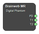
Brainweb MR
Class: NodePhantomBrainwebMR

This node is a tool for generating parameter maps relevant for magnetic resonance images, e.g. T1 and T2 maps. The node is based on the BrainWeb Phantom and two different brain datasets are available.
Example Workflows
Create T1, T2, T2* and PD maps
Outputs
PD [Optional output]
Proton density map.
Type: Image4DFloat
T1 [Optional output]
T1 map in units of seconds.
Type: Image4DFloat
T2 [Optional output]
T2 map in units of seconds.
Type: Image4DFloat
T2* [Optional output]
T2* map in units of seconds.
Type: Image4DFloat
X [Optional output]
Magnetic susceptibility map.
Type: Image4DFloat
CSF [Optional output]
Cerebrospinal fluid (CSF) map. Fraction of tissue in each pixel that is CSF.
Type: Image4DFloat
Skull [Optional output]
Skull tissue map. Fraction of tissue in each pixel that is skull.
Type: Image4DFloat
Dura Mater [Optional output]
Dura mater tissue map. Fraction of tissue in each pixel that is dura mater.
Type: Image4DFloat
Fat [Optional output]
Fat tissue map. Fraction of tissue in each pixel that is fat.
Type: Image4DFloat
Connective [Optional output]
Connective tissue map. Fraction of tissue in each pixel that is connective tissue.
Type: Image4DFloat
Bone Marrow [Optional output]
Bone marrow tissue map. Fraction of tissue in each pixel that is bone marrow.
Type: Image4DFloat
Muscles [Optional output]
Muscle tissue map. Fraction of tissue in each pixel that is muscle.
Type: Image4DFloat
Muscles Skin [Optional output]
Muscles or skin tissue map. Fraction of tissue in each pixel that is muscle or skin and not counted as muscle.
Type: Image4DFloat
Gray Matter [Optional output]
Gray matter map. Fraction of tissue in each pixel that is gray matter.
Type: Image4DFloat
White Matter [Optional output]
White matter map. Fraction of tissue in each pixel that is white matter.
Type: Image4DFloat
Vessels [Optional output]
Vessel map. Fraction of tissue in each pixel that are vessels.
Type: Image4DFloat
Background [Optional output]
Map indicating if a pixel is background.
Type: Image4DFloat
Settings
Phantom
Subject Selection
Select dataset from which the maps or masks should be generated.
Values: Subject04, Subject05
Tissue Parameters Text
Edit the tissue specific parameter values used in the image generation.
Source Tissues
Specify what tissues to be used when generating parameter maps. By default all tissues are selected.
CSF Boolean
Use CSF tissue for parameter maps generation.
Skull Boolean
Use skull tissue for parameter maps generation.
Dura Mater Boolean
Use dura mater tissue for parameter maps generation.
Fat Boolean
Use fat tissue for parameter maps generation.
Connective Boolean
Use connective tissue for parameter maps generation.
Bone Marrow Boolean
Use bone marrow tissue for parameter maps generation.
Muscles Boolean
Use muscle tissue for parameter maps generation.
Muscles Skin Boolean
Use muscle/skin tissue for parameter maps generation.
Gray Matter Boolean
Use gray matter for parameter maps generation.
White Matter Boolean
Use white matter for parameter maps generation.
Vessels Boolean
Use vessels for parameter maps generation.
Output Maps
Specify what type of parameter maps to generate.
PD Boolean
Enable proton density map output.
T1 [ms] Boolean
Enable T1 map output.
T2 [ms] Boolean
Enable T2 map output.
T2* [ms] Boolean
Enable T2* map output.
X Boolean
Enable susceptibility map output.
Ktrans [min⁻¹] Boolean
Enable Ktrans map output.
ve Boolean
Enable ve map output.
vp Boolean
Enable vp map output.
Output Tissues
Specify which tissue masks to generate.
CSF Boolean
Enable CSF tissue fraction map output.
Skull Boolean
Enable skull tissue fraction map output.
Dura Mater Boolean
Enable dura mater tissue fraction map output.
Fat Boolean
Enable fat fraction map output.
Connective Boolean
Enable connective tissue fraction map output.
Bone Marrow Boolean
Enable bone marrow tissue fraction map output.
Muscles Boolean
Enable muscles tissue fraction map output.
Muscles Skin Boolean
Enable muscles/skin tissue fraction map output.
Gray Matter Boolean
Enable gray matter fraction map output.
White Matter Boolean
Enable white matter fraction map output.
Vessels Boolean
Enable vessels fraction map output.
Output Masks
Background Boolean
Enable background map output.
Geometry
Settings determining the geometry of the generated maps.
Edit Boolean
Enable modification of geometry. Note: If not set, all geometry settings will be ignored and a default image size will be created.
Matrix X Integer
Number of pixels in x-direction.
Matrix Y Integer
Number of pixels in y-direction.
Matrix Z Integer
Number of pixels in z-direction.
Resolution X [mm] Number
Resolution in x-direction, i.e. the size (in mm) of a pixel in the x-direction.
Resolution Y [mm] Number
Resolution in y-direction, i.e. the size (in mm) of a pixel in the y-direction.
Resolution Z [mm] Number
Resolution in z-direction, i.e. the size (in mm) of a pixel in the z-direction.
Position offset RL [mm] Number
Image offset in the x-direction.
Position offset AP [mm] Number
Image offset in the y-direction.
Position offset SI [mm] Number
Image offset in the y-direction.
Rotation axis RL Number
Oblique slices can be obtained by rotating the image around an axis given in three coordinates in (RL, AP, SI) directions. This setting specifies the rotation axis component in RL-direction. The rotation vector does not need to be normalized.
Rotation axis AP Number
Oblique slices can be obtained by rotating the image around an axis given in three coordinates in (RL, AP, SI) directions. This setting specifies the rotation axis component in AP-direction. The rotation vector does not need to be normalized.
Rotation axis SI Number
Oblique slices can be obtained by rotating the image around an axis given in three coordinates in (RL, AP, SI) directions. This setting specifies the rotation axis component in SI-direction. The rotation vector does not need to be normalized.
Rotation Angle [degrees] Number
The angle to rotate the image around the rotation axis. See the setting: Rotation axis RL/AP/SI.
Extrapolation value Number
Value of voxels outside the phantom.
Interpolator Selection
Interpolator used when resampling data.
Values: NearestNeighbour, Linear, BSpline, Gaussian, BlackmanWindowedSinc, CosineWindowedSinc, HammingWindowedSinc, LanczosWindowedSinc, WelchWindowedSinc
References
1. http://www.bic.mni.mcgill.ca/brainweb/
2. C.A. Cocosco, V. Kollokian, R.K.-S. Kwan, A.C. Evans : "BrainWeb: Online Interface to a 3D MRI Simulated Brain Database" NeuroImage, vol.5, no.4, part 2 / 4, S425, 1997-- Proceedings of 3 - rd International Conference on Functional Mapping of the Human Brain, Copenhagen, May 1997.
3. R.K.-S. Kwan, A.C. Evans, G.B. Pike : "MRI simulation - based evaluation of image - processing and classification methods" IEEE Transactions on Medical Imaging. 18(11):1085 - 97, Nov 1999.
4. R.K.-S. Kwan, A.C. Evans, G.B. Pike : "An Extensible MRI Simulator for Post - Processing Evaluation" Visualization in Biomedical Computing(VBC'96). Lecture Notes in Computer Science, vol. 1131. Springer-Verlag, 1996. 135-140.
5. D.L. Collins, A.P. Zijdenbos, V. Kollokian, J.G. Sled, N.J. Kabani, C.J. Holmes, A.C. Evans : "Design and Construction of a Realistic Digital Brain Phantom" IEEE Transactions on Medical Imaging, vol.17, No.3, p.463--468, June 1998.
6. B. Aubert-Broche, D.L. Collins, A.C. Evans: "A new improved version of the realistic digital brain phantom" NeuroImage, in review - 2006.
7. B. Aubert-Broche, M. Griffin, G.B. Pike, A.C. Evans and D.L. Collins: "20 new digital brain phantoms for creation of validation image data bases" IEEE TMI, in review - 2006
See also
MRI Emulator BETA, Gradient-echo contrast BETA, Spin-echo contrast BETA
Keywords: BrainWeb, MRI parameter maps, Tissue maps
Copyright © 2022, NONPI Medical AB
