ISE
Class: NodeInducedSusceptibilityEffect

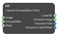
Produces the induced susceptibility effect for the supplied magnetic susceptibility map in form of the local B0-field, distorted image and distortion field. The input values are the image matrix together with the magnetic susceptibility map and patient mask.
Example Workflows
Induced susceptibility example
Inputs
Image
The image to be destorted due to the susceptibility effect.
Type: Image4DFloat, Required, Single
Susceptibility
The susceptibility map that generates a B0-field determening how Image will be destorted.
Type: Image4DFloat, Required, Single
Mask
Confine the distortion calculation to the region defined in the Mask.
Type: Image4DBool, Required, Single
Outputs
Local B0
The susceptiblity induced B0 field in units of Tesla.
Type: Image4DFloat
Distorted Image
The resulting distorted version of the input Image.
Type: Image4DFloat
Distortion Field
The distortion field in mm.
Type: Image4DVector3
Distortion Field Shifted
The distortion field in mm with the bulk avarege distortion removed.
Type: Image4DVector3
Settings
B0 Number
The field strength (T) of the B0-field
Lorenz Correction Boolean
Applies the Lorentz sphere correction
Positive Shift X Boolean
Sets the direction of the x-gradient in relation to the image matrix
Positive Shift Y Boolean
Sets the direction of the y-gradient in relation to the image matrix
Positive Shift Z Boolean
Sets the direction of the z-gradient in relation to the image matrix
Bandwidth X (Hz/Voxel) Number
The applied bandwidth (frequency encoding) in the X-direction given in Hz/Voxel
Bandwidth Y (Hz/Voxel) Number
The applied bandwidth (frequency encoding) in the Y-direction given in Hz/Voxel
Bandwidth Z (Hz/Voxel) Number
The applied bandwidth (read out) in the Z-direction given in Hz/Voxel
Gyromagnetic Ratio (Hz/T) Number
The gyromagnetic ratio to use. Typically this is the default value for protons of 42.57e+6 Hz/T
References
- J A Lundman, M Bylund, A Garpebring, C Thellenberg Karlsson, T Nyholm. Patient-induced susceptibility effects simulation in magnetic resonance imaging. Physics and Imaging in Radiation Oncology. (2017) DOI: 10.1016/j.phro.2017.02.004
- “The Insight Segmentation and Registration Toolkit” www.itk.org
See also
Keywords:
Copyright © 2022, NONPI Medical AB
