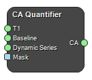
CA Quantifier
Class: NodeCAQuantifier
Calculates contrast agent (CA) concentration map based on a \(T_1\) map [ms] a baseline signal (signal at zero Ca concentration) and dynamic data acquired during which CA is present in the imaged region. All images must correspond to the same region and Baseline and Dynamic Series must have the same settings. An optional mask can also be provided to limit the calculations to a region.
The following signal models are supported:
Spoiled gradient echo
CA concentration is found from the ratio of the dynamic signal and the baseline signal by inverting the spoiled gradient echo equation(ignoring \(T_2^*\)-effects).
Saturation Recovery
CA concentration is found by using the signal equation
\(S=S_0(1-\exp(-T_I/T_1))\)
and a ratio between baseline (\(S_0\)) and dynamic signal (\(S\)). In the signal equation T1 is related to the contrast concentration \(C\) through
\(T_1^{-1} = T_{10}^{-1} + r_1C\)
In these two equations \(TI\) is the saturation delay (referred to as inversion time in the DICOM data) and \(T_{10}\) is the \(T_1\) without contrast agent.
Outputs a CA concentration map [mM].
Inputs
T1
Input T1 map.
Type: Image4DFloat, Required, Single
Baseline
Input baseline image.
Type: Image4DFloat, Required, Single
Dynamic Series
Input dynamic series.
Type: Image4DFloat, Required, Single
Mask
Input mask.
Type: Image4DBool, Optional, Single
Outputs
CA
Resulting contrast agent concentration map.
Type: Image4DFloat
Settings
Model Selection
Signal model.
Values: SPGR, SaturationRecovery
R1 (mM¯¹s¯¹) Number
T1 relaxivity of the CA.
Set Undefined Numbers To Number
Undefined values(failed fits) are set to this value.
References
- M.C. Schabel and D. L. Parker, “Uncertainty and bias in contrast concentration measurements using spoiled gradient echo pulse sequences,” Phys. Med. Biol., vol. 53, no. 9, pp. 2345–2373, May 2008
- Blüml Msc, Stefan & R. Schad, Lothar & Stepanow, Boris & J. Lorenz, Walter. (1993). Spin-lattice relaxation time measurment by means of a TurboFLASH technique. Magnetic Resonance in Medicine. 30. 289 - 295. 10.1002/mrm.1910300304.
Keywords: dce, dynamic, contrast, enhanced, t1, map
Copyright © 2022, NONPI Medical AB